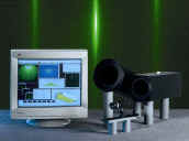
kSA 400 Imaging and Analysis System
HARDWARE FEATURES
Option of either digital or analog high-resolution, Peltier cooled CCD imager with software-selectable exposure time. 60 or 110 frames/second, 10-bit digital acquisition capability, 12 bit up-grades available.
Scientific-grade real-time image acquisition hardware, allowing real-time image display both on host computer monitor and optional auxiliary monitor.
Optimized optics for RHEED imaging, allowing for zoom in and zoom out without compromising light flux.
Flange mounting assemblies for a variety of vacuum chambers and flanges, concealing and protecting imager and optics and providing a light-tight environment.
Optional analog and digital input and output boards for reading external input (from thermocouple, for example) or controlling external devices (process control system, for example).
External triggering capability, including asynchronous reset for triggering precisely to external events, like substrate rotation. Includes fast shuttering up to 1/10,000 sec.
|
|
|
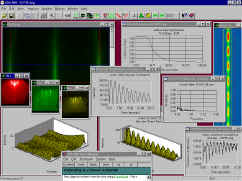
SOFTWARE FEATURES
Complete image archiving capability, storing image as well as image acquisition parameters (time, user comments, exposure time, image types, etc.)
Up to 96-bit deep image storage, resulting in higher accuracy for integration control and frame co-adding.
Real-time display of the following diffraction features (during growth, annealing, etc.):
- intensities (peak, average, centroid)
- lattice spacing
- FWHM/coherence
- line profile(s)
- analog input voltages
RHEED-specific analysis routines, including algorithms for determining in-plane lattice spacing, in-plane coherence length, and deposition rate.
Image "filters", including background subtraction, inelastic background subtraction, contrast maximization, high and low pass filters, false coloring, edge detection, 2D FFT, and more.
Optional user-programmable processing and analysis routines.
Various types of image acquisition, including single image, multiple images, multiple regions, and movies. "Scan Mode" images naturally show diffraction pattern evolution and yield simple time-resolved diffraction analysis. All images and graphics may be exported to standard file formats, including .wmf, .bmp, .tif, .avi, and text files.
Publication quality 2D and 3D graphics allow you to visualize diffraction data and fine-tune graphics by changing fonts, font sizes, labels, colors, palettes and axes.
Online, context-sensitive user manual.